The objective of the task
At the end of the nineteen-nineties a team of scientists from Oxford University in England determined the structure of the Isopenicillin N synthase enzyme, IPNS for short, which originates from the fungus Aspergillus Nidulans. The scientists observed that the active site of the enzyme is composed of three amino acids: two Histidine acids (marked with the letter H) and one Glutamic acid (marked with the letter D). These amino acids are located in different regions of the protein sequence at positions 214, 216 and 270 as seen in Figure 1.
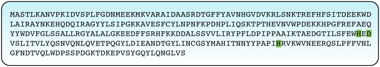
Figure 1: Sequence of the IPNS enzyme with the amino acids of the active site highlighted at positions 214, 216 and 270.
These findings raise two interesting research questions: how is it possible for two amino acids that are not adjacent on the sequence to create one active site? How does the substrate bind to the active site?
- How is it possible that the active site of the IPNS enzyme is composed of the amino acid Histidine at position 270, which is almost 60 amino acids away from positions 214 or 216, where the rest of the amino acids composing the active site are located? Is it due to the active site spreading over a large area of the enzyme?
Stages of the task
In order to answer this question we must remember the differences in the primary, secondary and tertiary structures of proteins. These differences will also help us understand how the active site is constructed and how the substrate binds to it. For this purpose in this activity we will perform the following steps:
- In this activity we will observe the spatial structure of the IPNS enzyme, which is also known as the 3D structure of the enzyme, by using a tool for displaying three dimensional structures of molecules called Jmol:
- We will take our first steps with Jmol and view the protein structure from various angles.
- We will display the secondary structure of the enzyme.
- We will identify the amino acids composing the active site and highlight them in the three-dimensional space. In this way we will be able to examine the spatial structure of the active site.
- We will observe how the substrate binds to the active site.