Identifying calcium ion binding regions
Calcium ions exist in the surrounding area of the living organism, and are significant to various processes in the cell. Scientists found that calcium ions are essential for stabilizing the structure of the MpAFP protein and for its function in preventing water from freezing.
13. To what level of organization do the calcium ions belong in the MpAFP protein?
Choose one answer:
- Primary structure – “linear structure” formed by the sequence of amino acids in the chain and the peptide bonds between them.
- Secondary structure – a structure in which hydrogen bonds are formed between certain amino acids causing the formation of one of two main structures: alpha helix or beta sheet (and turns and loops between them).
- Tertiary structure – a structure which gives the protein its ability to function and is created by various bonds (hydrophobic interactions, ionic interactions, hydrogen bonds, disulfide bridges (S-S) etc.).
- Calcium ions are not part of the protein structure.
The correct answer is D. Calcium ions have an important role in stabilizing the protein structure, but they do not constitute part of the protein itself. In addition to the structural stability provided by the calcium ions, it was also found that in their presence the protein acts as antifreeze and prevents ice from forming, and the loss or release of calcium ions from the protein would result in the loss of this function.
From a scientific standpoint, it is important to find where the calcium ions are located in relation to the entire subunit, and which areas in the protein bind the calcium ions. In order to more prominently mark the calcium ions, we will perform the following actions:
- To select the calcium ions (the element symbol is Ca) connected to subunit A only, open the console by right clicking the mouse and choosing Console in the additional options menu, and in the console window, right after the $ sign, type Select Ca:A and press Enter (thirteen atoms have been selected).
- To increase the volume of the calcium ions, right click the mouse in the workspace and pick:
Style ➔ Atoms ➔ 75% van der Waals - To change the color of the calcium ions to green, right click the mouse in the workspace and pick:
Color ➔ Atoms ➔ Green
Now it is easier to observe the calcium ions. You can see the calcium located between the loops (screen 12). At this stage it is recommended to rotate the structure in order to distinguish the relative location of the calcium ions in relation to the loops.
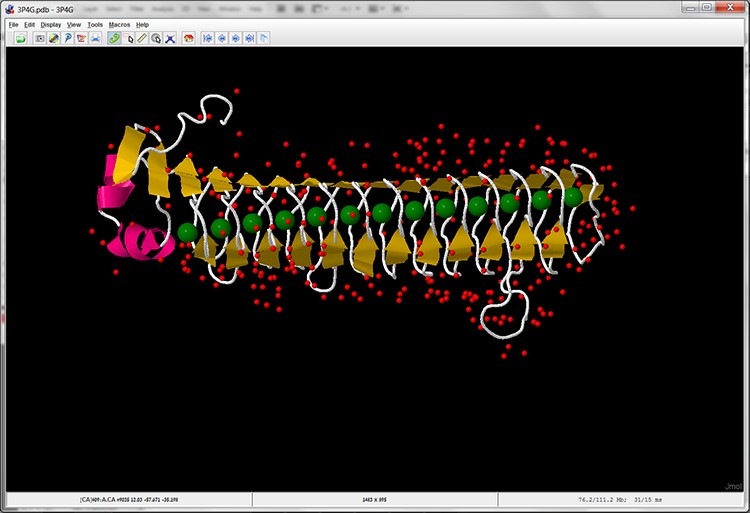
Screen 12: Cartoon display of subunit A of the MpAFP protein with calcium ions marked in green.
In order to learn more about the sequence characterizing the loops in the area binding the calcium ions, we will hide the water molecules in the protein structure model. For this purpose we will select the water molecule (in the additional options menu pick: Select ➔ Hetero ➔ All water)
And then we hide them, in the additional options menu pick (Style ➔ Atoms ➔ Off):